Cancer cells in motion
January 3, 2019
Dylan Burnette, PhD, assistant professor of Cell and Developmental Biology at Vanderbilt, uses a structured illumination microscope to capture cancer cells in motion. This advanced microscope shines different patterns of light on a sample to reveal images that are blurred by older microscope designs. The microscope he and other researchers use is housed at the Vanderbilt Cell Imaging Shared Resource, which is a Nikon Center of Excellence, one of only 21 such centers worldwide.
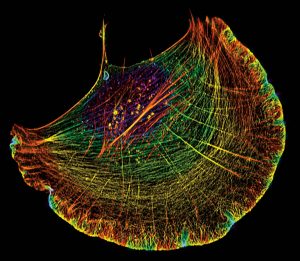
- The actin cytoskeleton shows a cell from an osteosarcoma. The cytoskeleton allows a cell to change its shape in order to move and is critical for human development, wound healing and the function of the immune system. However, when gone awry, the cytoskeleton can also drive the growth and metastasis of cancer.
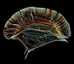
- The actin cytoskeleton in this crawling cell shows a normal fibroblast. The cytoskeleton allows a cell to change its shape in order to move and is critical for human development, wound healing and the function of the immune system. However, when gone awry, the cytoskeleton can also drive the growth and metastasis of cancer.
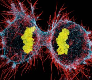
- A major way a tumor can grow is by cell division of cancer cells. This image shows an osteosarcoma cell that is about to divide into two new daughter cells. The cellular forces driving the separation of the two daughter cells are powered by two major components of the cytoskeleton: actin (red) and myosin II (blue). DNA, which functions as the cell’s genetic hard drive, is shown in yellow.
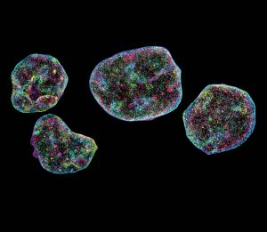
- DNA encodes the genetic material of each cell and is located in the cell’s nucleus. This image shows the 3D position of DNA in the nuclei of four cells isolated from an osteosarcoma.
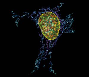
- This image features the powerhouses of our cells, mitochondria. These organelles, which look worm-shaped in this image, not only generate much of a cell’s chemical energy but also are hubs for cellular decision making, such as whether a cell lives or dies. This image shows the DNA in the nucleus.
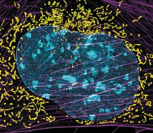
- This image features the powerhouses of our cells, mitochondria. These organelles, which look worm-shaped in this image, not only generate much of a cell’s chemical energy but also are hubs for cellular decision making, such as whether a cell lives or dies. This image shows the DNA in the nucleus and also shows the actin cytoskeleton (purple).
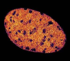
- This image, which somewhat resembles a chocolate chip cookie, is actually DNA within a single nucleus.

