Sound Power
New technology sends ultrasound waves to destroy tumors
January 14, 2025 | Paul Govern

In October 2023 the Food and Drug Administration approved a groundbreaking treatment for patients with liver cancer or liver metastases that uses focused ultrasound waves to destroy tumors. With philanthropic assistance, Vanderbilt University Medical
Center will begin to offer this innovative therapy, known as histotripsy, in early 2025.
Unlike surgery and other localized but invasive ablation treatments that rely on heat, cold or chemicals, histotripsy employs precisely targeted ultrasound waves to break down cancer cells through mechanical forces alone. The procedure typically takes half an hour and allows most patients to return home the same day.
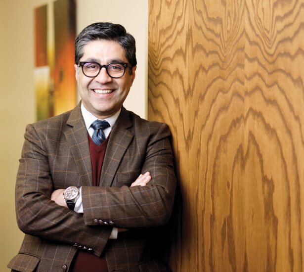
“A lot of patients with liver cancer or liver metastases are unfortunately not surgical candidates, and they run out of options. We can offer this treatment to selected patients, and what excites me is that it’s completely noninvasive,” said Kamran Idrees, MD, MSCI, MMHC, professor of Surgery and Ingram Associate Professor of Cancer Research, who is helping to lead the introduction of histotripsy at VUMC. He serves as chief of the Division of Surgical Oncology and Endocrine Surgery and director of Pancreatic and Gastrointestinal Surgical Oncology.
Histotripsy works by delivering microsecond pulses of high-pressure sound waves that create rapid pressure changes in tumor tissue. These changes cause microscopic bubbles to rapidly form, expand and collapse, generating intense and highly localized mechanical forces strong enough to break apart cancer cells. Beyond treatment for liver tumors, potential additional applications for histotripsy remain under study.

“The ultrasound waves go into the liver and create these small micro bubbles that oscillate and cause tissue disruption,” Idrees said. “It liquefies the tumor cells completely.” The destroyed tissue is naturally eliminated by the body within weeks to months.
A key advantage of histotripsy is its ability to preserve important anatomical structures while destroying cancer cells. “The best part about it is tissue selectivity,” Idrees said. “It affects the tumor cells, but it doesn’t affect the key structures within the liver such as blood vessels and the bile ducts.”
This selective destruction represents a significant advancement over current treatments for liver cancer, which include surgical removal, thermal ablation, chemoembolization and radiation therapy. While these traditional methods can be effective, they come with significant limitations. Surgery is invasive and suitable for only 10% to 20% of patients. Other treatments risk complications such as bleeding or injury to normal structures such as bile ducts. These traditional techniques will continue to have a role in treatment of liver tumors and liver metastases, Idrees said, with histotripsy figuring as a new addition to the armamentarium.
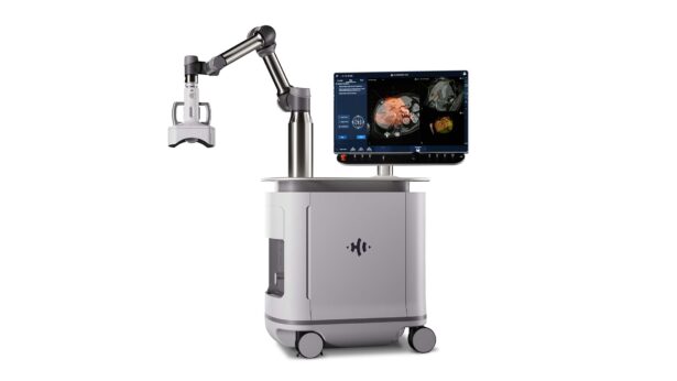
The procedure itself is remarkably straightforward. It’s performed under general anesthesia to control breathing, as the liver sits close to the diaphragm. Using ultrasound imaging guidance, physicians identify the tumor’s location and program these coordinates into a robotic system that delivers the treatment with precision. With a specialized membrane placed over part of the patient’s abdomen, a water bath between the membrane and the machine helps transmit the ultrasound waves effectively.
While the procedure can take as little as five minutes for smaller tumors, average treatment time per tumor is about 34 minutes.
The treatment can be monitored by doctors in real time using ultrasound imaging like what’s used for seeing babies in the womb.
“The patient comes in under general anesthesia; we identify the lesions under ultrasound; and we can ablate from a single lesion up to three lesions,” Idrees said. “After destruction of a lesion or lesions, we wake the patient up. If the patient is doing well, they go home the same day or after 23 hours observation. We cannot do that with any surgery or any other technology as of now.”
Another significant advantage is its repeatability for patients who develop new tumors over time. “It’s not one and done,” Idrees said. “If they have more lesions or other lesions pop up, we can repeat the procedure as needed.”
Unlike chemotherapy or radiation therapy, histotripsy doesn’t create heat or ions that could damage healthy DNA. The destroyed cancer tissue simply turns into a harmless liquid that gets naturally absorbed and eliminated by the body’s drainage systems.
One particularly notable advantage of histotripsy is that patients can continue their chemotherapy during treatment. As Idrees points out, “For any surgery and other procedures that we do, we stop chemotherapy for at least four to six weeks. Then they’re going to get the procedure, and if there’s no complication, they can restart the chemotherapy. But if they have a complication, there may be a delay in restarting the chemotherapy. With histotripsy, we don’t have to worry about any of that.”
And since the risk of bleeding with histotripsy is negligible, patients can continue blood thinners.
The treatment has shown promising results in clinical trials. In the HOPE4LIVER trial, which led to FDA approval, the technical success rate was 95%, with 42 out of 44 tumors successfully treated. The procedure-related major complication rate was 7%, which is comparable to existing treatments. The trial included patients with both primary liver cancer and liver metastases from other cancers, including colorectal, breast and pancreatic cancers.
The treatment can also be combined with other approaches. For patients with tumors on both sides of the liver, doctors can use histotripsy to treat one side while surgically removing tumors on the other side. Additionally, histotripsy has been approved for “downstaging” liver tumors — reducing their size to make patients eligible for liver transplantation.
Idrees notes that, regarding the liver, histotripsy does have some current limitations; notably, the liver lesion must be visualized by ultrasound and the maximum treatment depth is 14 to 16 centimeters, making tumors near the top of the liver and certain right-sided liver lesions difficult to access and treat.
While currently approved only for liver tumors, research is ongoing to explore histotripsy’s potential use in treating other types of cancer. The technology’s ability to preserve healthy tissue while effectively destroying tumors makes it a promising addition to the cancer treatment arsenal.
