Virtual mapping for facial reconstruction
December 6, 2016

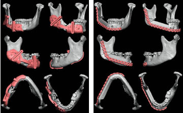
Three-dimensional CT scans are used with virtual surgical planning (VSP) to develop guides for facial reconstruction. The photo on the left depicts the cutting guide design, while the one on the right pinpoints the final position of plates.
In the past, traditional craniofacial reconstruction allowed little pre-planning; surgeons had to decide exactly where they would cut while they were in the operating room. Now, through the use of virtual surgical planning (VSP), physicians can plot out surgeries in advance.
One of the first steps in a facial reconstruction is to run computed tomography (CT) scans of the area; the CT scans can then be used to create 3D images that provide a clear, spatial view of head and neck structures. These views can be rotated and studied by the plastic surgeon so every precise cut of bone and tissue and every placement of plates and screws can be mapped.
In the 1990s, 3D models of the skull based on a patient’s CT scans began being used for planning reconstructive surgeries. Now, a further dimension has been added to this process. Plastic surgeons at Vanderbilt send CAT scans to companies that create cutting “jigs” or guides that show precisely where cuts need to be made on the section of bone being removed and on the bone being relocated to repair the defect.
“In the past, it was almost like we were in the woodshop out back,” said Wes Thayer, M.D., Ph.D. “You’d measure, and then you’d cut. There is definitely an art to it in that you improve your techniques with time, but we’ve compared all of our surgeries in the plastic surgery department before we used computer-based modeling to after, and the improvement in surgery time was both significant and in fact, remarkable.”
On average, the plastic surgeons spend three hours and 45 minutes less time in surgery, per case, when they use virtual surgery planning because they are able to accurately fit the pieces together more quickly to make the repairs, Thayer said.
