The MDS challenge
The molecular maze of bone marrow failure
June 2, 2016 | Bill Snyder
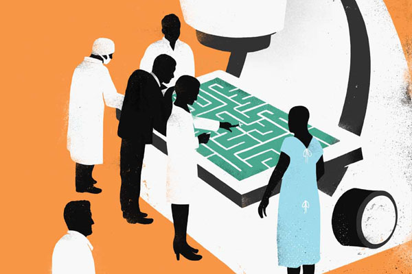
Illustration by Sébastien Thibault
Each year at least 45,000 Americans experience bone marrow failure from a variety of disorders known as myelodysplastic syndromes, or MDS.
They lose the ability to manufacture healthy blood cells that are needed to prevent infection or even carry out simple clotting. Something goes awry in the bone marrow, where blood cells are produced, but what exactly causes these disruptions remains a mystery.
Among older Americans, MDS is more common than melanoma and considerably more lethal. About one-third of patients will develop acute myeloid leukemia (AML), a fast-growing cancer of bone marrow cells. “Most are gone within a few years, or less,” said Michael Savona, M.D., director of Hematology Research at the Vanderbilt-Ingram Cancer Center (VICC).
Standard treatment options are limited to supportive care, disease-modifying drug therapy, or chemotherapy with stem cell transplantation, the only method that offers the possibility of a cure.
Vanderbilt researchers are determined to turn that picture around. Thanks to cutting-edge science and a highly collaborative culture, they are mapping out the molecular labyrinth of MDS and exploring new approaches to halt, prevent or even reverse bone marrow failure.
Their efforts have not gone unnoticed. Last year, Vanderbilt University Medical Center announced a research partnership with Incyte Corporation to study the therapeutic potential of some of its compounds for treating MDS and other cancers.
Grants from the National Institutes of Health and from EvansMDS, an initiative of the Edward P. Evans Foundation, are helping Vanderbilt scientists explore at the cellular, molecular and genetic levels what goes wrong when the bone marrow fails. The MDS Foundation has designated VUMC a Center of Excellence.
“Learning from the science to help people quickly—that’s really our mission,” said Savona, principal investigator of the Incyte grant and director of the VICC Hematology Early Therapeutics Program.
Teams of VICC scientists are working on multiple fronts to better understand MDS and develop new treatments.
Why cells die
According to Sandra Zinkel, M.D., Ph.D., myelodysplastic syndromes are not failure of the bone marrow to produce blood cells. Rather, she said, “as fast as they’re made, they’re dying … They don’t get out into the blood.”
Zinkel, associate professor of Medicine, Cancer Biology, and Cell and Developmental Biology, joined forces in 2012 with Carlos F. Lopez, Ph.D., a systems biologist newly recruited from Harvard, to figure out why cells in the bone marrow unexplainably die in MDS.
There are two forms of “programmed” cell death. One form, called “apoptosis,” is normally used to remove cells that are no longer needed. Cells simply “implode” and the leftovers are recycled by the immune system as part of cellular housekeeping.
Necrosis, the other major form of cell death, triggers an immune system alarm bell, in contrast to the silent apoptosis process. Cells that encounter a viral invader, for example, will “explode.” The cellular “shrapnel” they release sets off an inflammatory immune response to fight off the invading virus.
Curiously, MDS cells in the bone marrow die preferentially by necrosis, rather than apoptosis. Zinkel and her colleagues have found that unrestrained programmed necrosis in a mouse model leads to bone marrow failure closely resembling human disease.
It’s not known why this happens, but three proteins—BID, Rip kinase and caspase-8—play a role.
BID is the acronym for a protein that guides cells down the silent, apoptosis pathway. Mice lacking BID develop a fatal disorder that closely resembles a form of human leukemia.
Rip kinase and caspase-8 are opposing enzymes—the first promotes necrosis while the second helps switch on apoptosis. In MDS patients, bone marrow cell death is caused by increased necrosis signaling through Rip kinases.
Drugs that inhibit Rip kinases potentially could reverse this process. But first, researchers have to understand the forking paths in this cell-death labyrinth.
To do that, Lopez developed computational models based on the expression levels and activity of the three proteins in the context of programmed cell death, and used the nation’s largest supercomputers at Tennessee’s Oak Ridge National Laboratory to carry out his simulations.
His models suggested that targeting a single protein in isolation would not sufficiently redirect the path from necrosis to apoptosis. Instead, it appeared that interactions between the three proteins determined whether the cell imploded or exploded.
“Focusing on one protein alone is like aiming at a moving target,” said Lopez, assistant professor of Medicine, Biomedical Engineering and Biomedical Informatics, “but understanding the mechanism of complex formation means we could instead ambush them.”
The right splice
Another researcher, Melanie Ohi, Ph.D., is exploring the spliceosome.
The human genome is made up of about 25,000 genes. And yet it encodes approximately 90,000 distinct proteins. How can that be?
Meet the spliceosome, a macromolecular “machine” that, with greater accuracy than an automated jigsaw, cuts pieces out of pre-messenger RNA and patches the new ends together to create mature messenger RNA, which is then translated into protein. Different splices, different proteins.
Sometimes, however, the machine goes awry. More than half of all patients with MDS have mutations in spliceosome components. Ohi is studying mutations in the spliceosomes of the blood cells in MDS patients that render their jigsaws worse than useless.
“These are spontaneous mutations,” said Ohi, associate professor of Cell and Developmental Biology. “My hypothesis is that (mutated) spliceosomes … splice incorrectly. Some of the genes they splice incorrectly go on to actually function as a protein and mess up signaling pathways.”
This is not uncommon: 30 percent of MDS patients have a mutation in one of the protein parts of the spliceosome called SF3B1.
To find out how the mutation affects the function of the spliceosome, Ohi uses a technique called single particle cryo-electron microscopy. Spliceosomes, either with or without MDS mutations, are biochemically purified, frozen and imaged using an electron microscope. Computational techniques are applied to the images to determine the three-dimensional structure.
Once Ohi and her colleagues have determined the structure of a spliceosome, “we have a pretty good idea where everything is,” she says.
They may, for example, be able to predict how a patient’s mutation will affect function of the spliceosome, even if that mutation has never been seen before. “That’s pretty powerful,” Ohi said. “It’s personalized structural biology, in a sense.”
Drug-like molecules have been developed that target the spliceosome, and one actually binds to SF3B1, the component that is mutated in many MDS patients.
“I don’t think this has been tested in the clinic yet, but the idea is that if you treat patients with a spliceosome inhibitor, maybe the MDS cancer cells would be hit harder than normal cells because the spliceosome is already compromised and that would be enough to kill (them),” Ohi said.
“You’d have to be careful, because the spliceosome is important for all cells,” she added. “It’s a fine line.”
Decoding the protein riddle
“Reading” the genetic code is a highly regulated process.
Misregulation and mutation of transcription factors, which are proteins involved in that process, can lead to misreading of the DNA—and cancer. One example is Myc, a family of transcription factors that is overexpressed in the majority of malignancies and contributes to an estimated 100,000 cancer-related deaths each year in the United States.
Last year, Bill Tansey, Ph.D., and colleagues discovered how Myc may work—through a cleft in a chromosome-binding protein called WDR5.
By slipping a “loop” of itself in the WDR5 crevice, Myc turns on genes involved in growth and development, said Tansey, Ingram Professor of Cancer Research. In the case of cancer, this leads to runaway growth.
In collaboration with Tansey’s group, a team led by Stephen Fesik, Ph.D., solved the crystal structure of the Myc-WDR5 interaction. Now they’re testing small molecules for their ability to block the crevice and prevent Myc from binding to the DNA.
“That’s what keeps me awake at night,” said Shaun Stauffer, Ph.D., associate director of medicinal chemistry in the Chemical Synthesis Core, part of the Vanderbilt Institute of Chemical Biology. “How are we going to prevent Myc from binding to DNA?”
Fesik, the Orrin H. Ingram II Professor of Cancer Research, came to Vanderbilt from Abbott Laboratories, where he developed a method for screening small chemical fragments for their ability to bind to “pockets” on the surfaces of proteins.
He and his colleagues use nuclear magnetic resonance or X-ray crystallography to determine how the binding occurs. This information can show them how to link the fragments into compounds with potential drug-like activity.
Last year, in addition to WDR5, the Tansey-Fesik collaboration turned up a protein called host cell factor 1 (HCF1). It binds to the mysterious central portion of Myc, a highly conserved and therefore presumably essential region of the protein present in all vertebrates.
HCF1 is a very abundant protein, in part because it binds to two other proteins, ASXL1 and BAP1, which stabilize it and prevent it from being destroyed (recycled) by the cell.
In a significant number of MDS patients, however, especially in those who progress to AML, there are mutations in one of the two proteins.
“The hypothesis that we’re testing is that when you lose ASXL1 or BAP1 … HCF is cleared from the cell and this works to alter the function of Myc,” Tansey said.
A global reach
Knowledge shared is knowledge advanced.
That’s the unofficial motto of the MDS effort at Vanderbilt, which increasingly has a national and even global reach. EvansMDS, the Evans Foundation initiative that is supporting a half-dozen Vanderbilt projects, will hold its annual research summit at VICC in October.
The latest research findings about MDS also are included in My Cancer Genome, launched by VICC in 2011 as the first online precision cancer medicine knowledge resource.
It is one of the projects supported by EvansMDS.
Today My Cancer Genome covers 22 different malignancies and cancer-associated disorders like MDS, and more than 300 genetic mutations. A globally shared resource for research, the site logs more than 9,000 visits a week.
Of molecules and mice
The first drug to treat MDS was approved in 2006. Since then, a few more drugs have been approved for various forms of the disease, but the progress has been frustratingly slow.
It can cost over $1 billion and take 15 years to bring a new drug to market. That’s why biopharmaceutical companies like Incyte increasingly are partnering with academic medical centers like VUMC to do high-risk discovery research.
For example, Jonathan Irish, Ph.D., assistant professor of Cancer Biology, uses a complex array of antibodies generated against proteins inside and outside the cell, a technique called single-cell phenotyping, to determine which proteins within each cell type are affected by compounds developed at Incyte.
Savona, principal investigator of the Vanderbilt-Incyte Alliance grant, grows myeloid disease from patient samples in genetically engineered, humanized mice and tests with various new drug therapies and combination therapies. He collaborates with Irish to determine what cell types are affected by the drugs and how the drugs affect cellular signaling. Those that make the cut may go into Phase I clinical testing in patients.
“We have three or four drugs in Phase I right now which will be important for the next generation of therapies for MDS patients,” he said. “We aim to help provide the right therapies to the appropriate patients as quickly as possible.”