3D Ingenuity
Surgeon develops visual aid for real-time pathology consults
November 30, 2023
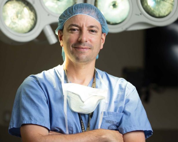
Three-dimensional scanning and printing systems are now utilized for purposes ranging from same-day dental crowns to prosthetic limbs, so Michael Topf, MD, wondered why computer-aided design (CAD) technology hadn’t made it into operating rooms as a visual aid for real-time consultations.
Surgeons communicate via phone and are dependent upon verbal descriptions from pathologists for guidance about resection margins. Those real-time, intraoperative consultations about how much more tissue should be cut to remove any lingering cancer cells are a critical component of surgical oncology. Pathologists scrutinize resected tumors, which are flash frozen and taken to their labs for microscopic analysis, while the surgeons await the results in the operating room.
“Frequently when a cancer specimen is removed, sectioned and processed, it is destroyed,” said Topf, assistant professor of Otolaryngology-Head and Neck Surgery. “Sometimes, two-dimensional photographs are taken of that specimen, but usually we have no visual record of the resected cancer specimen. When a question comes up about a particular margin or how something was processed, we rely on verbal communication without any visual aid between the surgeon and pathologist to clarify that margin location.”
Since no protocol was available for this purpose, Topf decided to create it. It took him and his team two years. A top priority was developing a protocol that didn’t significantly interfere with workflow.
“We know that surgeons are already pressed for time and that pathologists and the pathology lab have to process a ton of specimens,” he said. “We couldn’t come up with a protocol that was going to interrupt or interfere in any way. That would never be adopted even if the information generated by that protocol is worthwhile. We came up with a way to 3D scan a surgical specimen in real time in less than 10 minutes prior to processing and not interfere with all the other important things that are going on in the lab.”
The 10 minutes, however, is not the total time required. Collectively, it takes about an hour, but only 10 minutes are added to the frozen section analysis process. Surgeons and pathologists who have adopted the protocol report that it’s beneficial. A survey of early adopters, which was published online Sept. 22, 2002, in the Head & Neck journal, showed that 85.8% of surgical team members and 74.9% of pathology team members reported that the system achieved clearer communication.
Communication is especially important with surgeries for head and neck cancer because positive margin rates with oral cavity squamous cell carcinoma are the highest among solid tumor malignancies. However, the visual aid can be used with other cancer surgeries.
“It would be applicable to any cancer surgery that removes cancer in an en bloc fashion, which is most cancer surgeries,” Topf said. “We have done 3D scanning for orthopaedic oncology surgeons and for breast cancer surgeons. It is applicable to most solid malignancies. It is widely transferable.”
The system was developed using easily accessible parts, but Topf and his team are looking to upgrade some components.
“Thus far, with the 3D scanning, we have used commercially available 3D scanners that you can buy on Amazon,” he said. “The one we are using now is a 3D scanner. It’s a middle-of-the-road 3D scanner and commercially available CAD software. We are interested in developing our own software for annotation of models. We have intentionally kept this relatively simple. Therefore, it is widely transferable to other institutions. We have gone to one other institution and shown them how to do 3D scanning.”
The 3D renditions of the excised tumors of individual patients are another important contribution to the image library for personalized care.
“This is taking the CT scan, the MRI scan or a PET scan and adding to that a 3D model of an actual cancer tumor,” he said. “It’s improving the whole cancer care team’s understanding of that patient’s disease process.”
The need for such an image may seem obvious, but there are reasons why the concept hasn’t been executed.
“All of this was right there in front of us,” Topf said. “Part of the reason for the lack of adoption and no one else wanting to do this has to do with, that as surgeons and pathologists, we are very busy. We process a lot of specimens. We perform a lot of surgeries. We thought we didn’t have the time to add another thing to the surgical/pathology workflow. We spent a lot of time developing the protocol, so that it got down to a very finite window that did not interfere with the process. Surgeons typically don’t like change. They are creatures of habit. People are resistant to change, particularly when they are busy.”
His 3D research initiatives have received support from a Discovery Grant from the VICC Ambassadors and a P3 Catalyst Award.
It is not yet known whether the imaging system will improve surgical outcomes. That will depend on the results of future prospective studies comparing results utilizing 3D imaging of pathology specimens versus standard of care and verbal communications. But Topf said he believes it will improve cancer care.
“They say a picture is worth a thousand words,” he said. “I don’t know how many words a 3D specimen model is worth, but it is certainly worth a lot.” n